Sonoembryology
Sonoembryology
The embryonic period begins at the moment of fertilization and continues until the 10th week of gestation. During this time, the embryo is very small, and its anatomical structures undergo rapid and significant changes. The introduction of diagnostic ultrasound (sonography) into clinical practice has enabled the safe and non-invasive assessment of the embryo during early pregnancy. High-frequency transvaginal ultrasound allows for an even closer and more detailed examination of embryonic morphology and development.
Sonoembryology is a specialized branch of sonographic obstetrics focused on the systematic evaluation of the anatomy and developmental stages of the human embryo. The term sonoembryology was introduced in the 1990s to highlight the close correlation between ultrasound images and actual embryonic development.
Cardiac activity in the embryo is usually detectable after the completion of 6+0 weeks of gestation. In the subsequent weeks, the initial development of the central nervous system, spine, limbs, stomach, and the physiological midgut herniation (omphalocele) becomes visible. The advent of 3D ultrasound and advanced software applications has greatly enhanced the quality of ultrasound images, making the early detection of abnormal embryonic development more feasible.
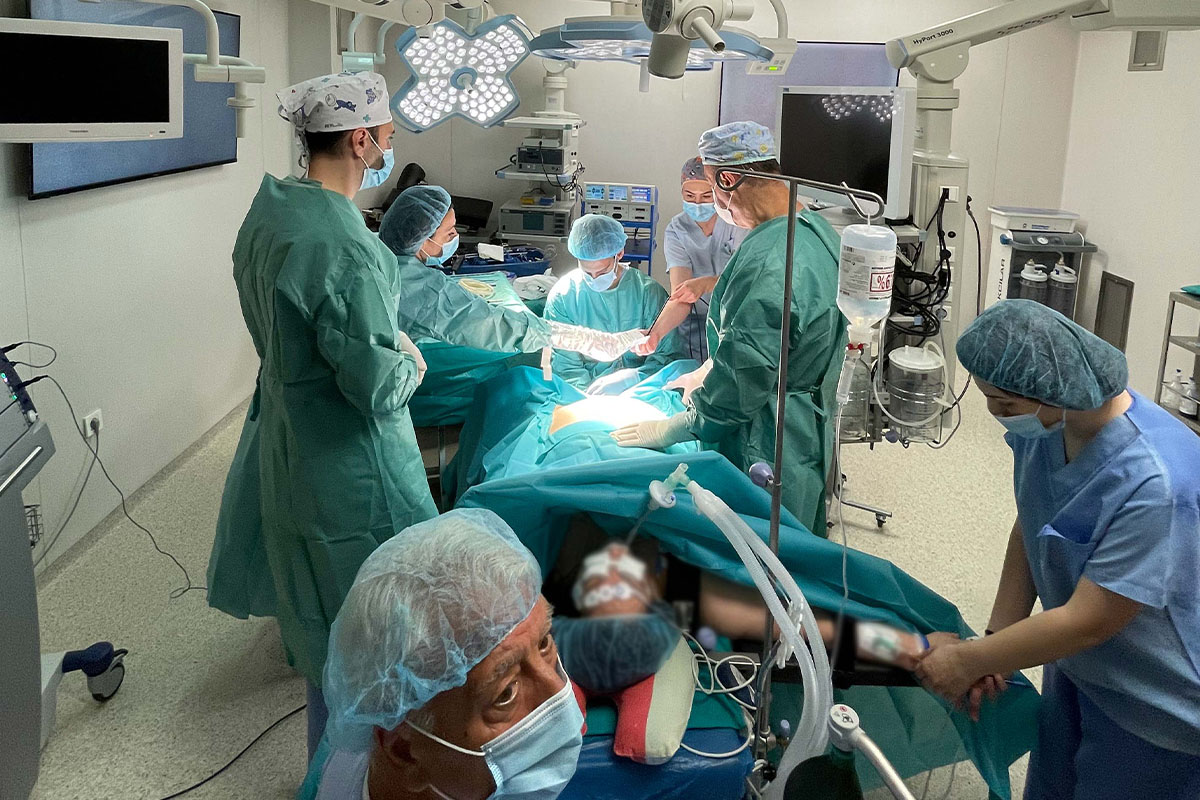
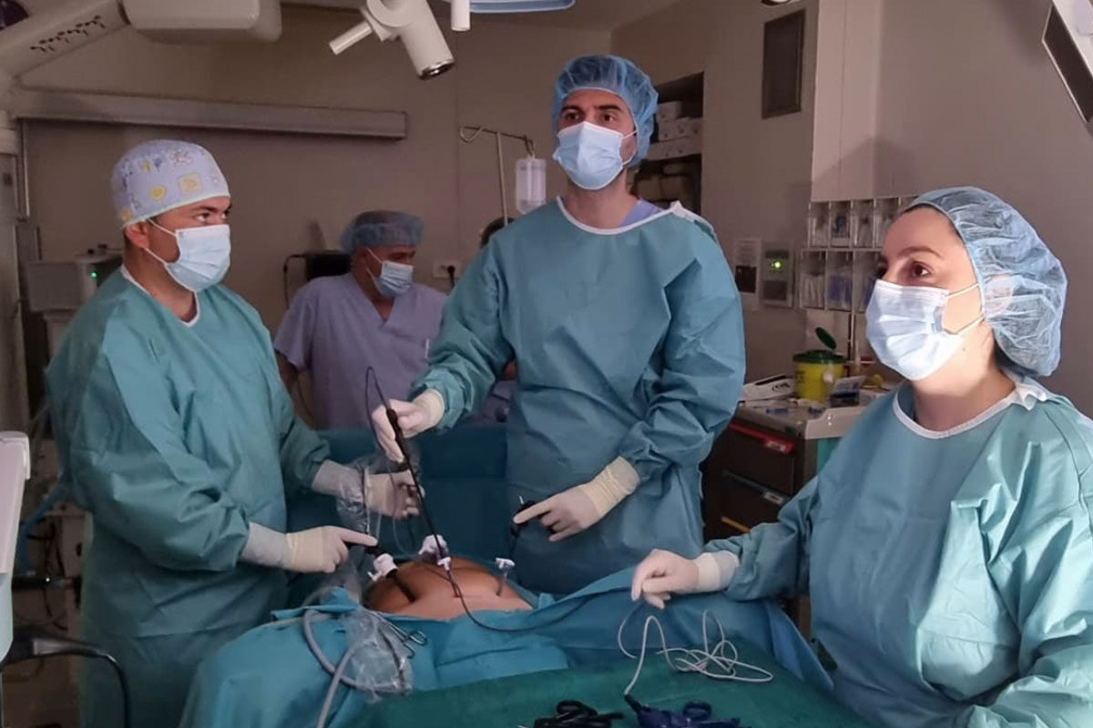
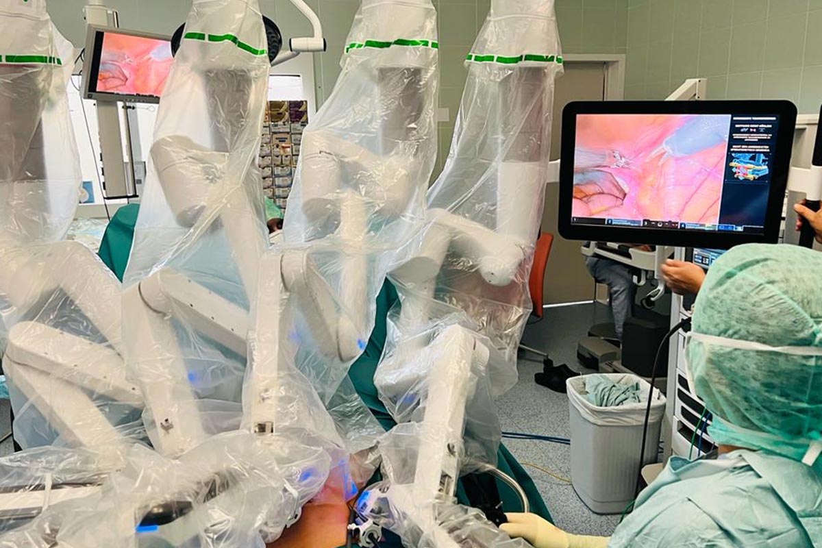
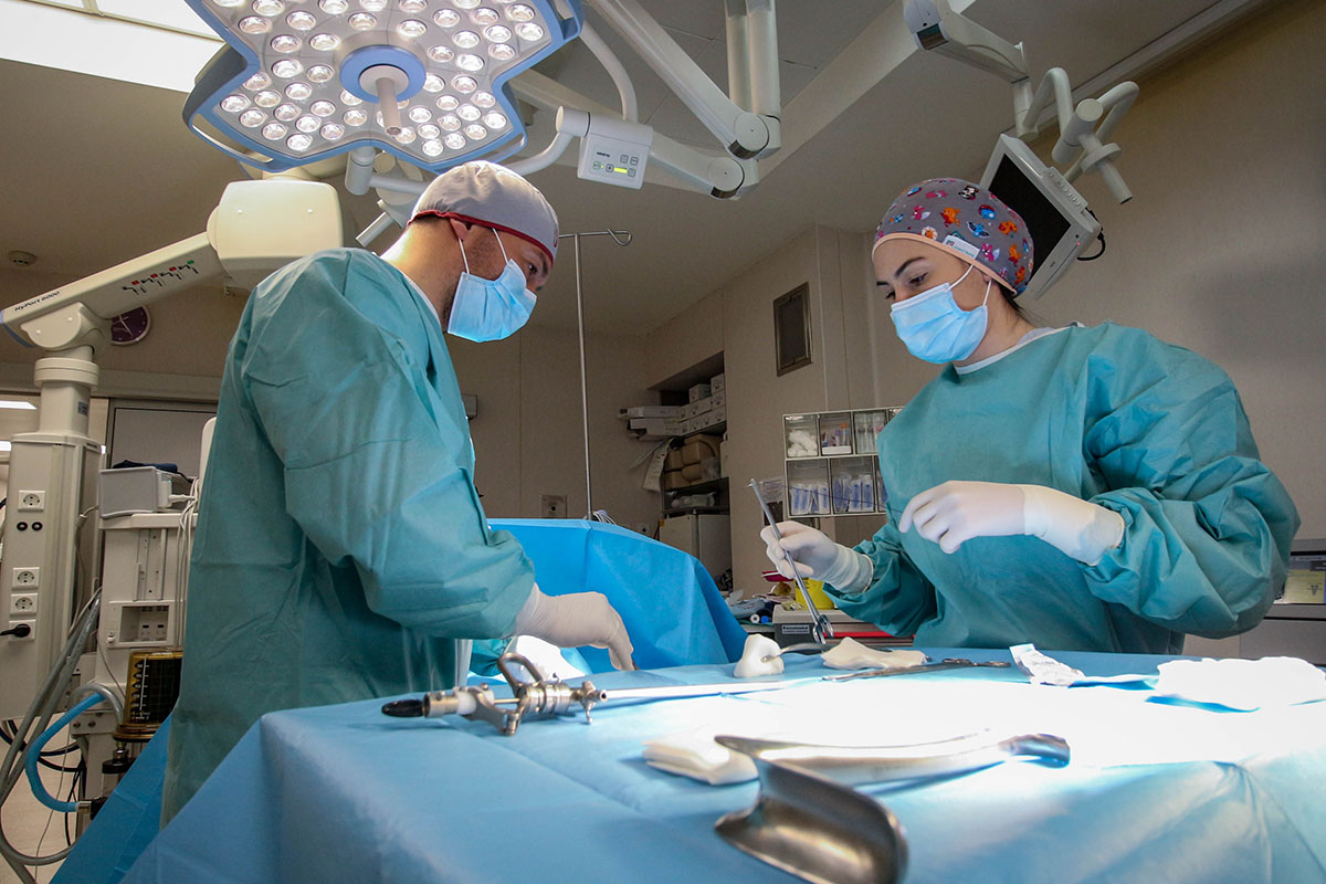
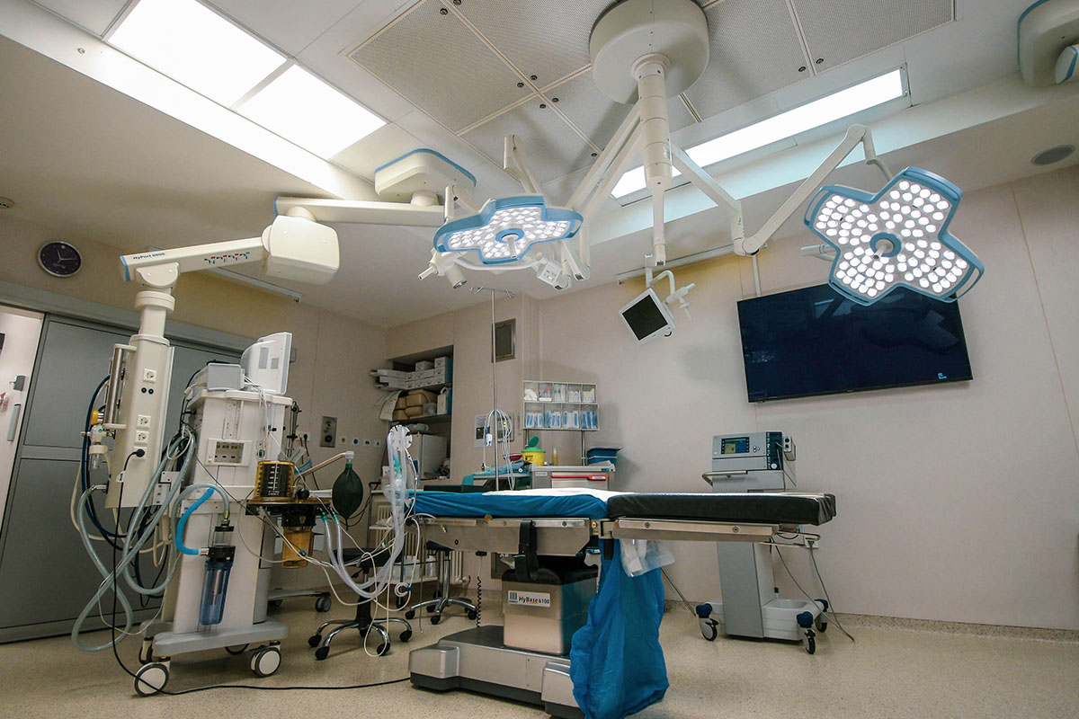
At Hospital Plodnost, we have the latest ultrasound equipment and highly qualified specialists who can monitor your pregnancy from the earliest stages. For more information or to schedule an appointment, please consult your doctor or contact our registration desk.


Gynaecology and Obstetrics Doctors

Prim. D-r Tashe Trpchevski
Consultant Gynaecologist and Sub-Specialist in Reproductive Medicine and Surgery

D-r Hristijan Trpchevski
Specialist Gynaecologist & Obstetrician

D-r Vladimir Popovski
Specialist Gynaecologist & Obstetrician

D-r Blagojce Obednikovski
Specialist Gynaecologist & Obstetrician

D-r Kristinka Pajakovska
Specialist Gynaecologist & Obstetrician

D-r Marija Hristovska
Specialist Gynaecologist & Obstetrician

D-r Vladko Gjorgjievski
Specialist Gynaecologist & Obstetrician

D-r Sotir Ropi
Specialist Gynaecologist & Obstetrician

D-r Aleksandar Jovanovski
Specialist Gynaecologist & Obstetrician

D-r Ana Vangelov
Specialist Gynaecologist & Obstetrician

D-r Sanja Milunovikj
Trainee Specialist Gynaecologist & Obstetrician
Medical-Surgical Nurses

Emilija Srbinoska
Responsible Medical Surgical Nurse

Meri Mihevska
Medical Nurse

Natasha Momiroska
Medical Nurse

Zaneta Veselica-Stefanovska
Medical Nurse

Maja Miloshevska
Medical Nurse

Biljana Dimitrovska
Medical Nurse

Sonja Aleksovska
Medical Nurse

Nikolina Risteska
Medical Nurse
Medical Nurses Midwifes

Merlinda Drala
Responsible nurse, gynecology and obstetrics department

Nadica Neshkovski
Medical Nurse

Jasmina Bozinovska
Medical Nurse

Aleksandra Dimovska
Medical Nurse

Fiona Kelja
Medical Nurse

Irena Talevska
Medical Nurse

Hristina Bumbaroska
Medical Nurse

Martina Karanfilovska
Medical Nurse

Dijana Boshevska
Medical Nurse

Sonja Fuzevska
Medical Nurse

Zaneta Vasilevska
Medical Nurse
Contact Us
SAT 08:00 – 13:00
Recent Posts

29 August 2025

29 August 2025
