Fetal Echocardiography
Fetal echocardiography, or Fetal Echo, is a specialized ultrasound examination focused on the fetal heart. Performed by our qualified Ob&Gyn (Fetal Medicine) specialists or pediatric cardiologists and we use advanced ultrasound technology. This examination is essential for the early detection of congenital heart defects (CHDs) which are among the most common fetal structural abnormalities, occurring in 8 to 12 per 1000 live births. Approximately half of these defects are severe, posing significant risks to life and often requiring surgery after birth. The incidence of CHDs during prenatal life is even higher, with a fetus being 5 to 6 times more likely to have a cardiac malformation than Down syndrome. Therefore, fetal echocardiography is the most effective method to exclude major congenital heart defects prenatally.
Fetal echocardiography can be performed at various stages of pregnancy. The expertise of our healthcare professionals and the quality of the ultrasound equipment we use are crucial factors in obtaining accurate results.
At Hospital Plodnost, we have a dedicated team of pediatric cardiologists trained specifically in fetal echocardiography. If a congenital heart defect is detected, a multidisciplinary team of experts thoroughly investigates the complex anatomy, hemodynamics, and prognosis.
Fetal Echocardiography at 12-13+6 Weeks Gestation:
- This examination can be conducted as early as 12-13+6 weeks, either transvaginally or transabdominally.
- Transvaginal fetal echocardiography offers significantly better resolution and image quality.
- Early fetal echocardiography at 12-13+6 weeks is indicated for high-risk pregnancies, such as those with a family history of congenital heart defects, gestational diabetes, or exposure to teratogens.
- Increased fetal nuchal translucency at this stage is another indication for early fetal echocardiography.
- The examination can also provide early reassurance to parents.
Caution: Due to the early gestational age and rapid development of the fetal heart, it is recommended that fetal echocardiography be repeated later in pregnancy.
Fetal Echocardiography Between 18-23 Weeks of Gestation:
- The fetal heart is still small and underdeveloped in the first trimester; therefore, fetal echocardiography is traditionally scheduled between 18-23 weeks (optimally at 20-22 weeks).
- During this period, a thorough assessment of the complex anatomy of the fetal heart is performed.
- In some cases, fetal echocardiography may also be required later in pregnancy, typically at 28-32 weeks.
For more information about the possibilities of having fetal echocardiography at Hospital Plodnost at any gestational age, please consult your doctor or contact our registration desk.


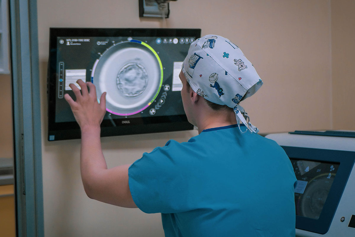
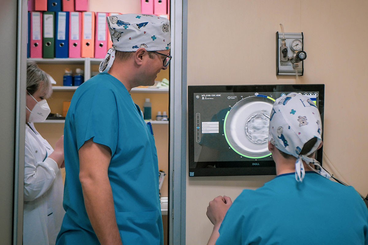
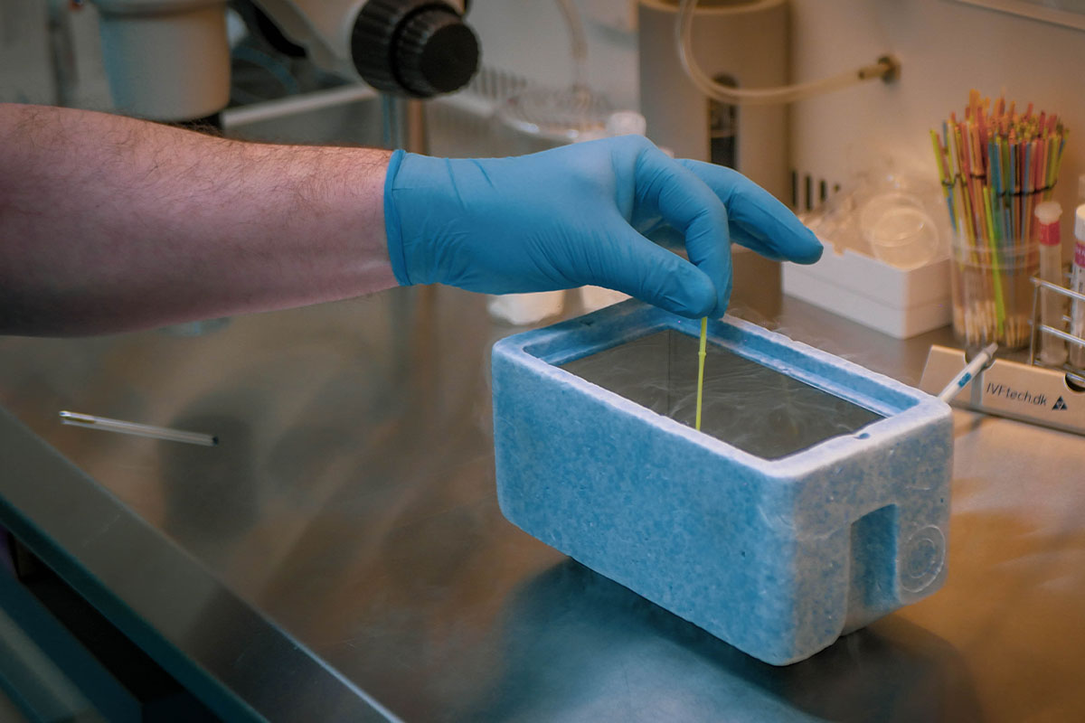
In-Vitro Fertilization (IVF) Department

Prim. D-r Tashe Trpchevski
Consultant Gynaecologist and Sub-Specialist in Reproductive Medicine and Surgery

Dr Nagja Trpchevska
Specialist Gynaecologist & Obstetrician

Goran Stefanovski
Clinical Embryologist-Andrologist
Prof. of Biology

M-r Stefan Matik
Clinical Embryologist-Andrologist
Master in Pharmacology (MPharm)

Nikolche Ognenovski
Clinical Embryologist-Andrologist
Biology Engineer

Sneshka Dimitrovic
Responsible nurse of the IVF department
Bachelor in Nursing

Aneta Joshevska-Damchevska
Medical Nurse
Contact Us
SAT 08:00 – 13:00
Recent Posts

29 August 2025

29 August 2025
